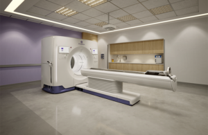
Powered by its proprietary Deep Silicon detector technology, GE HealthCare’s Photonova Spectra offers a flexible photon-counting computed tomography (PCCT) platform with multiple configurations to meet diverse clinical needs.
Chicago-based GE HealthCare says the system offers wide coverage and ultra-high definition (UHD) spacial and spectral imaging. It helps enable fast acquisition speeds and the precise visualization of subtle tissue variations, small lesions and vascular structures.
Unlike conventional CT converting X-rays into light before measuring, photon-counting CT directly counts individual X-ray photons. It measures their energy, enabling the potential for higher spectral and spacial resolution and improved tissue characterization. Deep Silicon, a novel detector material, enhances spectrial imaging, supporting advanced lesion characterization and treatment monitoring.
GE HealthCare submitted Photonova Spectra with advanced AI applications to the FDA in November 2025.
“As clinicians across the United States face rising volumes and increasing diagnostic complexity, technology must do more than capture images; it must simplify decision-making and strengthen performance across the enterprise,” said Catherine Estrampes, President & CEO, U.S. and Canada, GE HealthCare. “Photonova Spectra is designed to deliver rich clinical insights in every scan and help alleviate cognitive burden for care teams. With the U.S. 510(k) clearance, we are proud to now bring this innovation to U.S. healthcare systems and the patients they serve.”
More about the new GE HealthCare imaging technology
GE HealthCare says that its Deep Silicon feature with 8-bin energy resolution supports advanced material separation and characterization capabilities. It enables Photonova Spectra to clearly distinguish between different materials like iodine, calcium and fat with precision. Wide detector coverage and rapid rotation speed (0.23 seconds) also support fast acquisition and motion-free imaging.
Photonova Spectra automatically captures both 8-bin spectral and ultra-high definition spatial data simultaneously. It achieves this without special setup or protocols, delivering access to spectral information in every exam.
GE HealthCare says its system can enhance treatments in spaces like neurology, oncology, musculoskeletal imaging, thoracic imaging and cardiology. It can deliver visualization of tiny structures in the brain for neurology and characterize lesions in oncology. The system can visualize small fractures and bone marrow edema as well. Additionally, it offers ultra-high-definition chest scans and full chest imaging to enable in-stent lumen assessment, plaque characterization and myocardial assessment.
The company said its system can harness up to 50 times more data than conventional CT through Nvidia computing technology as well.
GE HealthCare also has ongoing collaborations with UW-Madison and Stanford Medicine to explore novel clinical applications and imaging protocols with Photonova Spectra’s advanced architecture.
The company plans to begin preparing for commercial availability in the U.S. with its FDA clearance secured.
“Photonova Spectra reflects years of intentional design and close collaboration with clinicians, researchers and collaborators across the globe,” adds Jean-Luc Procaccini, president & CEO, Molecular Imaging and Computed Tomography, GE HealthCare. “From the earliest stages to today, we remain focused on building a system that addresses the practical realities of clinical practice while opening pathways for scientific advancement. The result is a photon-counting platform engineered for the needs of today’s care teams, as well as the imaging challenges and research opportunities that will shape the future of CT.”